 “Leptospires are aerobic spirochetes whose cells are flexulous, motile, tightly coiled and have axial flagellae. Some are pathogenic, though others are harmless freshwater saprophytes”
- aerobic: requiring oxygen (dissolved in water) to survive.
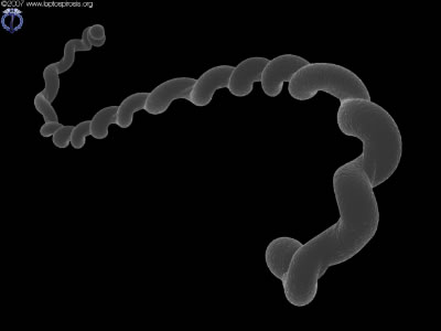
- spirochetes: a phylum of 6 bacterial genera, long and coily in shape (as in the photo).
- flexulous: able to bend and wriggle.
- motile: Able to propel themselves about.
- axial flagellae: a pair of rod-like structures around which the bacteria is ‘coiled’, which allows the bacteria to move by turning and wriggling. There is one from each end, both pointing towards the center.
- pathogenic: capable of causing illnesses.
- saprophytes: creatures that feed from decaying organic material.
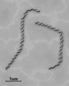
The bacteria are in general about 0.1µm in diameter and 10-20µm in length. In comparison, a red blood cell is about 7µm in diameter, so despite being quite long, the very small width of leptospires makes them difficult to see under optical microscopes unless a contrast-enhancing technique such as dark-field is used. They are gram negative and there is no visual difference between serovars. Live specimens move extremely quickly, and under visible light their rapid rotation can make them appear as a chain of dots instead of a continual structure. They are propelled by two flagella, one rooted to each end of the bacterium and inserted within the membrane, along the axis. The result looks like the bacterium has been wrapped around a stick, as in the photo. Virulent specimens usually have a characteristic ‘hooked’ shape but straight versions are known to exist. When damaged by chemicals or ingested by phagocytes, the bacterium can collapse into a tiny spherical ball, where it is presumed to have no pathogenic ability. Leptonema are generally larger (20% wider and 30% longer), and Turneria parva are smaller (about half the size). The spiral coiling of all leptospires is right-handed (the same as the thread on a bolt).
The ability of leptospires to move rapidy (up to 15µm per second) through water is vital to their life cycle, as they must spread out as much as possible to maximise the chance of infecting a new host. Their motion does not seem to help them penetrate tissues but it does impact on their ability to enter the bloodstream. Typically they spin rapidly on their axis, with one or both ends hooked – though when moving ‘forward’ the front end is often straightened. There is of course no ability to plan a route nor is there a defined front and back end, and they will move either way.
Classification is based on serological analysis of the antigens of leptospires and divides all of the bacteria into one of ten genomospecies. Each of these is divided into serogroups, and then within each group are the serovars. There are over 200 known serovars at this time, but only a fraction of them cause the human and animal illnesses which all fall under the term ‘leptospirosis’. The majority of known serovars are harmless organisms, or are only known to infect non-human hosts. The pathogenic bacteria are almost entirely within the L. interrogans genomospecies, and in that is the serogroup best known for human infection, Icterohaemorrhagiae. Worth noting is that a genomospecies has been named after Dr Weil (L. weilii) but this has no connection to Weil’s Disease, for which the cuplrit is L. interrogans serovar Icterohaemorrhagiae or Pomona. The classification and grouping of the bacteria can seem confusing in the least, and the species interrogans was actually named because of the number of questions being debated at the time!
 The image on the left is a computer-enhanced electron micrograph of leptospira – click on it for a large version. This image is copyright-free and you can use it however you wish.